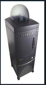
The IVIS Spectrum is an optical imager used to facilitate non-invasive longitudinal monitoring of disease progression, cell trafficking and gene expression patterns in living animals. Filters and spectral un-mixing algorithms let you take full advantage of bioluminescent and fluorescent reporters across the blue to near infrared wavelength region. It also offers single-view 3D tomography for both fluorescent and bioluminescent reporters that can be analyzed in an anatomical context using a Digital Mouse Atlas.
For advanced fluorescence imaging, the IVIS Spectrum has the capability to use either trans-illumination (from the bottom) or epi-illumination (from the top) to illuminate in vivo fluorescent sources. 3D diffuse fluorescence tomography can be performed to determine source localization and concentration using the combination of structured light and trans illumination fluorescent images. The instrument is equipped with 10 narrow band excitation filters (30nm bandwidth) and 18 narrow band emission filters (20nm bandwidth) that assist in significantly reducing autofluorescence by the spectral scanning of filters and the use of spectral unmixing algorithms. In addition, the spectral unmixing tools allow the researcher to separate signals from multiple fluorescent reporters within the same animal.
The instrument is located on the first floor of LKS. The instrument is suitable for animals (mice) and certain plant and microbial specimens.