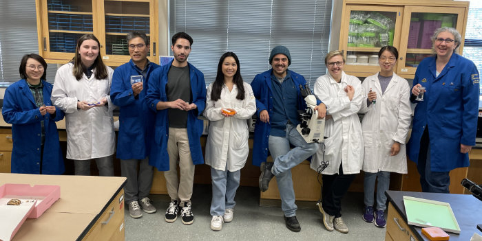
This year we had 8 students who learned plant & animal microtechnique
Plant and Animal Microtechnique 2025. Lead instructor Dr. Denise Schichnes on the right.
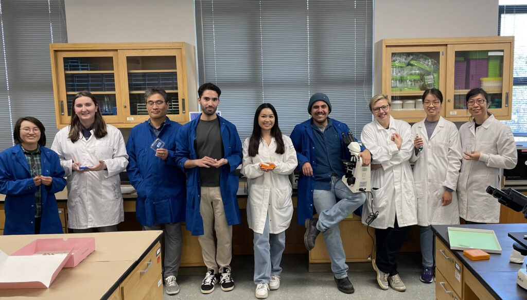
Plant and Animal Microtechnique June 2025. (Co-Instructor Dr. Jules Cho on the right.)
Examples of their work
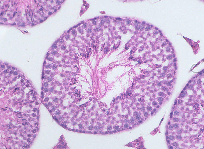
Cross section through seminiferous tubule in mouse testis, stained with Mayer’s Hematoxylin and Eosin. Imaged using 40x objective. Sample by Lourenço Martins.
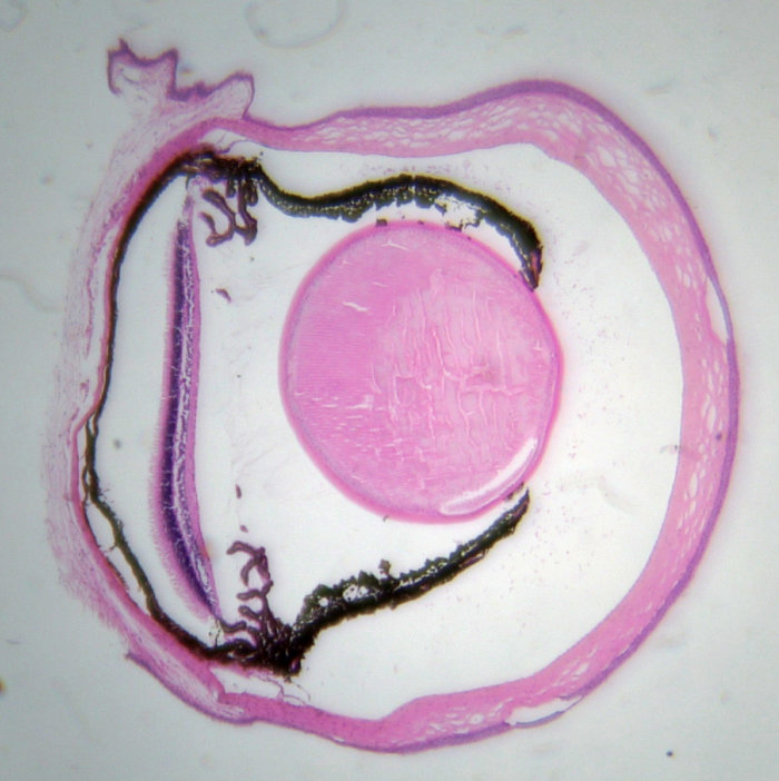
Cross section through mouse eye stained with Mayer’s Hematoxylin and Eosin. Imaged using 4x objective. Sample by Aimee Kiang.
Longisection of redwood meristem stained with Johansen’s Safranin O and Fast Green. Imaged with 4x objective. Sample by Sasha Nikolaeva.
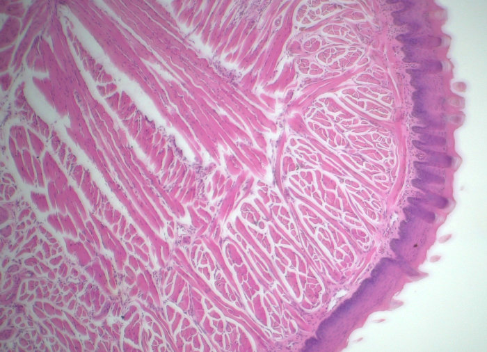
Mouse tongue stained with hematoxylin and eosin imaged using 10x objective. Notice papillae at margins. Sample by Y. Justin Choi
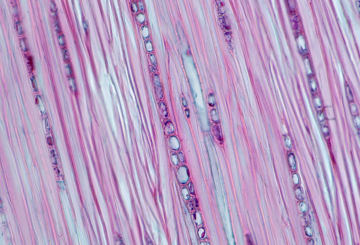
Rays in cross section of Mongolian Oak stained with Johansen’s Safranin O and Fast Green FCF. Imaged using 40x objective. Sample by Emanuele Guglielmini.
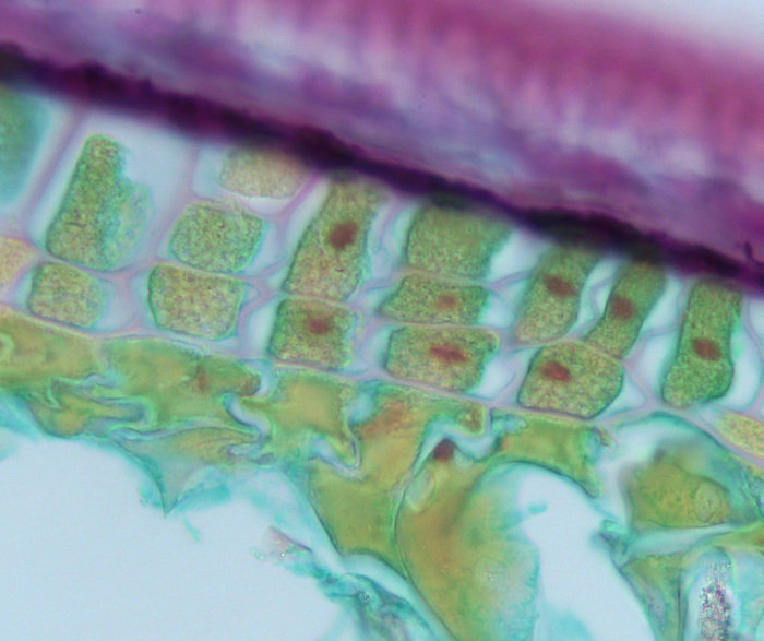
Cross section through Barley aleurone layer of a softened seed, stained with Johansen’s Safranin O and Fast Green. Imaged using 100x objective. Sample by Vanessa Bowman.
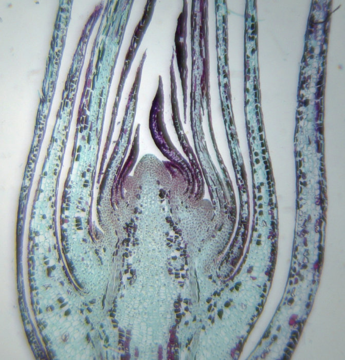
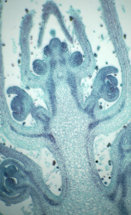
Longisection through a shoot apical meristem of Ocimum basilicum and stained with Johansen’s Safranin O and Fast Green. Imaged using 10x objective. Sample by Isabella Massaro.
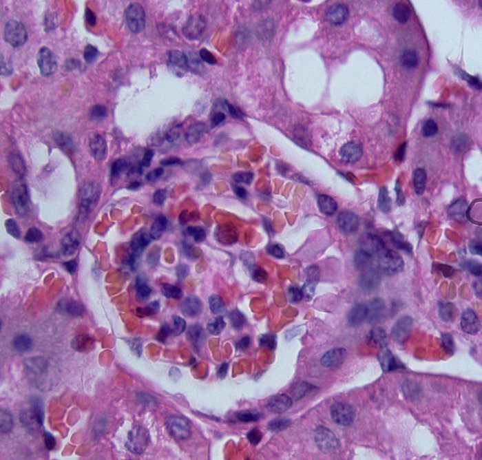
Section through a mouse kidney showing a glomerulus stained with Mayer’s Hematoxylin and Eosin. Imaged using 100x objective. Sample by Lan Mai.