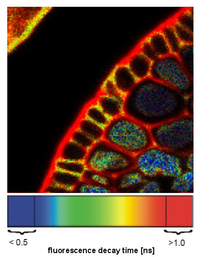
Fluorescence lifetime Imaging is a technique whereby a microscopic sample is imaged using a scanning fluorescence microscope (WF or confocal) to detect and measure the decay rate, or fluorescence lifetime, of fluorescent molecules within the sample. The goal is to initiate fluorescence, then measure decay of photon emission, as opposed to imaging the total fluorescence.
Thus, such an instrument has certain hardware requirements:
- The Ex light source may be a pulse laser or a rapidly changing sine wave of excitation (some from an LED), as opposed to a continuous illumination (from a lamp or CW laser). The system will measure fluorescence lifetime between laser pulses. Pulse widths are in the fs (femptosecond) range, with pulse frequency around 80 MHz.
- Since Fluorescence Lifetime is in the nanosecond range, fluorescence detectors must be extremely sensitive and fast.
- Additional electronics and detectors.

This is an example of a FLIM image. Coloration is pseudocolor. It relates to the fluorescence lifetime (in ns) and not to actual fluorescence emission spectra. This image shows, for a single fluorophore, that different environments within the root transverse section result in different fluorescence lifetimes. Cytoplasm generally decreases FL, whereas the cell wall environment increases FL.
Follow this link to see what a FLIM scope looks like.
What does FLIM tell us?
- What fluorophore is in the sample. This info is limited to the sample (pixel) size.
- What is the state of the environment (pH, Ca+2). FL changes relative to some environmental parameters.
- Is FRET occuring? FL changes if FRET happens.