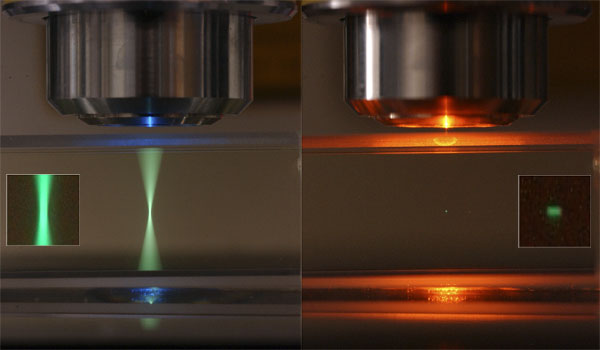
Image by Steve Ruzin and Holly Aaron, UC Berkeley
With 2P however, it's a little confusing. While it is still true that resolution is dependent on the wavelength of emitted light, the size of the excitation spot is so much larger than its equivalent in 1P confocal (i.e., in 1P it is not the resolution-limiting factor, in 2P it is) that the constant in the resolution equation is greatly increased. What limits resolution in all microscopy is spot diameter, and that diameter is dependent on the longest wavelength. In 1P the longer wavelength is the Em light. In 2P the longest wavelength is the Ex light.
Thus, for 2P resolution should be described as d=0.7(lambda-em)/NAobj. However, in this instance resolution is dependent on the non-linear two-photon effect, which occurs in a volume that is SMALLER than this equation predicts. Thus, given everything else equal, the Lateral and Axial resolution of 2P is equivalent to confocal imaging.